
You might be wondering, what is biological therapy? Well, it is a treatment that stimulates the immune system to fight diseases, infections, or even the side effects of some treatments.
0 Comments
What´s the truth behind popular diets such as the carnivore one?
Gestational trophoblastic disease (GTD) is a term used for a group of pregnancy related tumors that are rare. They begin in the layer of cells called the trophoblast that normally surrounds an embryo. Early in normal development, the cells of the trophoblast form tiny, finger-like projections known as villi. The villi grow into the lining of the uterus. In time, the trophoblast layer develops into the placenta, the organ that protects and nourishes the growing fetus. The main types of GTD are Hydatidiform mole (complete or partial), Invasive mole, Choriocarcinoma, Placental-site trophoblastic tumor and Epithelioid trophoblastic tumor.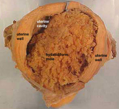 La enfermedad trofoblástica maligna es un tumor que se produce a partir de la degeneración maligna de la placenta. La mayoría de los casos aparecen tras un embarazo molar, en el que la estructura del feto se sustituye por un enorme racimo de pequeños quistes llenos de un líquido acuoso con pocas proteínas. En otras ocasiones, la mola es parcial y comparte un espacio con restos fetales inmaduros. Finalmente existen casos originados en un parto aparentemente normal, con un feto en perfecto estado. Afortunadamente, este origen es el menos frecuente, ya que en ocasiones, el tumor maligno no manifiesta hasta bastantes meses después del parto, dificultando sobre manera el disgnósitco, precisamente por la normalidad absoluta asociada al parto más o menos reciente. 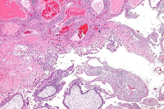 Acrogeria or Gottron´s syndrome is a skin condition characterized by premature aging, especially in the form of rare fragile skin on the hands and feet (distal extremities). It´s one of the most common congenital premature aging syndromes ocurring early in life and it was charaterized in 1940. In early childhood, it progresses over the years and remains stable over time with morphology and colour. It has been observed that there is a bruising tendency and it may affect mroe females than males. The causes are still not determined but the disorder is thought to be inherited as an autosomal recessive genetic trait. Mutations in the COL3A1 gene have been reported in varied phenotypes, including acrogeria and vascular rupture in Ehlers-Danlos syndrome (especially type IV). Some of the main signs and symptoms are loss of subcutaneous fat and collagen of hands and feet, skin lesions (can be associated with osteoarticular alterations), small hands and feet with unusually prominent veins, short stature and sometimes abnormally small jaw (micrognathia). It was originally described by Gottron in 1941 when he noticed premature cutaneous aging on the distal extremities in two brothers which were present since birth. It is extremely rare (40 reported caases in ,edical literature since 1941) and there is no especific treatment available. La acrogeria o síndrome de Gottron es una condición dermatológica caracterizada por un envejecimiento prematuro, especialmente en forma de una inusual y frágil piel en manos y pies. Es uno de los síndromes de envejecimiento prematuro congénito que ocurren en la vida temprana y qu fue descrito y caracterizado en 1940 por primera vez. En los primeros años de la infancia, progresa a lo largo de los años y se mantiene estable en el tiempo en cuanto a morfología y color. Se ha observado que hay una tendencia de formación de cardenales que probablemente afecte a más mujeres que hombres. Las causas todavía no están determinadas pero se cree que se hereda como un rasgo genético autosómico recesivo. Mutaciones en el gen COL3A1 se han alegado en varios fenotipos, incluyendo acrogeria y las rupturas vasculares en el síndrome de Ehlers-Danlos del tipo IV especialmente. Algunos de los signos y síntomas más comunes son: pérdida de grasa y colágeno subcutáneos de manos y pies, lesiones en la piel (pueden ir asociadas con alteraciones osteoarticulares), manos pequeñas y pues con inusiales venas prominentes, baja estatura y a veces una mandíbula anormalmente pequeña (micrognatia). Fue descrito por primera vez en 1941 por Gottron cuando se dió cuenta de un envejecimiento cutáneo prematuro en las manos y los pies de dos hermanos que habían estado rpesentes desde el nacimiento. Es una enfermedad muy rara (40 casos citados en la literatura médica desde 1942) y no hay un tratamiento específico para tratarla.A stye is a small abscess (painful collection of pus) on the eyelid and it is an infection at the root of an eyelash. They are very common and a person may have one or two during their lifetime. Styes are usually caused by an infection with staphylococcus bacteria. They appear as a small painful red lump, often with a yellow spot in the middle, on the outside of the eyelid. Other symptoms are watery eye and a red eye or eyelid. It´s not always necessary to see a doctor, although if you have a very swollen eyelid with a stye it should be checked. If you have been diagnosed with long-term blepharitis (inflamation of the eyelids) this may also increase the risk of styes. Most of them get better without treatment within a few days or weeks. They may burst and release pus after 3-4 days. A cloth warmed with warm water held against the eye encourages the stye to release pus and heal more quickly. Further treatment is not usually needed unless you have a very painful stye that is very swollen and that indicates spreading infection. In this case, the doctor may decide to treat it with antibiotics, drain it or refer you to an ophthalmologist.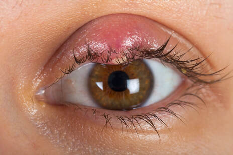 Un orzuelo es un pequeño absceso localizado superficialmente en alguna glándula /de Zeiss o de Moll) en la base de las pestañas. Son muy comunes y una persona puede llegar a tener de media un par de ellos en toda su vida. Los orzuelos suelen estar causados por una infección de staphylococcus, una bacteria. Aparecen como un pequeño y doloroso bulto rojo, a veces con un punto amarillo en el medio, en el exterior del párpado. Otros síntomas son los ojos llorosos y párpado u ojo rojo. No siempre es necesario ir al médico, pero si el párpado con el orzuelo están muy hinchados sí. Si te han diagnosticado blefaritis de larga duración (inflamación de los párpados), hay más probabilidades de aparición de orzuelos. La mayoría de ellos se pueden curar sin ningún tratamiento en varios días o semanas. Pueden estallar y expulsar pus en unos 3 o 4 días. Si se aplica una compresa caliente sobre el orzuelo, este acelera la expulsión de pus y se cura más rápido. No se suele necesitar más tratamiento al menos que se tenga un orzuelo muy doloroso e hinchado que indique esparcimiento de la infección. En este caso, el doctor lo más probable es que recete un antibiótico, lo drene o derivarte a un especialista en oftalmologíasIt´s a surgical procedure to treat congestive heart failure caused by myocardial infarction (heart attack). A heart attack, scar or an aneurysm may develop resulting in an enlarged rounded heart. This may lead to congestive heart failure (CHF). This surgery is done to restore the heart to a more normal size and shape, therefore improving function. The SVR procedure is usually performed in conjunction with coronary artery bypass grafting (CABG) to ensure optimal blood supply to the heart. Some patients will also have valve repair. To determine is a patient needs this surgery, we need to do an echocardiogram (ECHO) (a test which uses sound waves to create a picture of your heart and help us determine how well your heart functions as a pump), a cardiac Catheterization (Cath) (a catheter placed in your groin allows pictures to be taken using X-rays and dye. These pictures help your team determine the function of your heart, along with the condition of the coronary vessels of your heart, to determine if bypass grafting is needed) and a magnetic Resonance Image (MRI) (a test which uses magnetic imaging to create a picture of your heart and its structures, and helps guide surgical decision making. This is often a preferred way to determine if you are a candidate for SVR. If you have an AICD or pacemaker a special cardiac CT may be obtained). If tests have been performed recently, than they will not need to be repeated. The ECHO and or MRI will also be repeated 3 months and one year after your SVR to track the success of the procedure. It consists on placing the patient on a heart lung machine at first. Then the surgeon will make a small incision into the left bottom part of your heart, through scarred tissue. The heart is opened, and a plastic model of a specific size is used to reshape the heart. |
“Wherever the art of Medicine is loved, there is also a love of Humanity. ” 
|